© Pint of Science, 2025. All rights reserved.
Heart Anatomy is a collaboration between the scientist Catrin Rutland and the artists Deborah Gardner, Jess Parry, Rachel Rea, 4 women of different routes, age, and backgrounds.
Rachel studied Costume Design and Textiles. Later working professionally in film productions and events, she has since built upon and utilized her skills for her more recent projects in creating artistic sculptures and installations.
Jess is an award-winning Welsh artist. She recently graduated from her Fine Art degree in 2020, and currently is working towards her first ever solo show in the summer of 2021.
Deborah’s practice is process and materially led, ranging from oblique references to the body, place and embodied memory and structures, which refer to the multiple and the cellular. She is also a programme leader in Art & Design at the University of Leeds and a member of the Royal Society of Sculptors and LAND2 (Practice led research network).
Catrin is an Associate Professor of Anatomy and Developmental Genetics, Faculty of Medicine & Health Sciences at the University of Nottingham.
Her research concentrates on cardiovascular disorders (heart disease and angiogenesis) and anatomy. In her own words:
'I call my research ‘Mending a broken heart’. I look into differences in the heart, whether that is anatomical or genetics, to understand different heart conditions. One of our discoveries was a heart bone (os cordis) being present in chimpanzees with heart disease. My group also work on a heart disorder called dilated cardiomyopathy where the heart gets larger which can cause very serious heart problems including cardiac arrest. Researching the hearts of people and animals to understand disorders and anomalies, in order to try and find solutions, is hard work but very rewarding.'
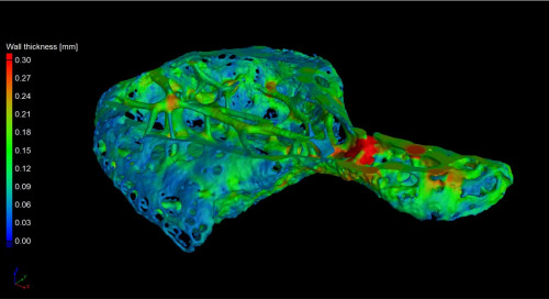
Delving into the Os Cordis, X-ray micro CT image, imaged using VGStudioMax, taken by Dr Craig Sturrock and Dr Catrin Rutland.
She also shared with us an inspiring story or event throughout her research career.
'I was once given permission by the Pope and the Vatican to study some rare anatomy books in the Vatican library for my research. I was nervous (don’t touch the books!) but seeing the original works by Da Vinci really inspired me to think beyond pure science and out into the world of science communication. Science communication is about informing other scientists and clinicians but it is essential to make our work available in different ways, to make it more accessible. SciComm was not a concept/career in Da Vinci’s day, but he certainly succeeded in a magnificent way.'
'One way in which I try to show young people the joys of science is via scientific papers, which include art, for young people (not so young people often love them too). For more information on hearts, anatomy, biology please do see my webpage with links to those papers. For those interested in the research side, you can see a list of my papers and research biography here.
'Much of the creative work has revolved around our os cordis discovery presented in this paper and this press release. If you are interested in cardiomyopathy, this paper and this paper for young people may be of interest.'
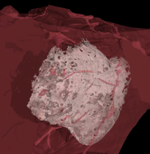
Os cordis within the chimpanzee heart tissue using computed micro-tomography and imaging reconstruction
The meetings between the artists and Catrin started immediately after being introduced to one another. Catrin shared her research with the artists to get an understanding of her projects and to start building their ideas. Catrin:
‘We discussed heart disease and the heart bone, the techniques I use in my scientific discoveries and even the scientific imaging I use to help uncover how the heart works and why. We talked a lot about how science can be portrayed, different techniques they use to reveal anatomy, structure and scientific concepts. We also talked about the emotions, feelings, and ethics behind undertaking scientific discoveries. As a scientist we rarely discuss the emotions behind science, yet these do come across in the brilliant pieces created by the artists.‘
Deborah Gardner
Deborah (in her own words) talks about her collaboration:
'In discussion of our three approaches to the discovery of the os cordis bone in a chimpanzee’s heart, I was struck by the variation of approach; Jess’s use of paint and the visceral living heart, Rachel’s dissection of the cow’s heart and the resulting layered images of her discoveries and my exploration of the bone ramped up massively in scale to the original of only a few millimetres. Using screenshots from Dr Catrin Rutland’s video footage scanning the bone, I rotated the image and used the infrastructure to explore branch-like networks to convey ideas of a tree of life. In filming my sculpture, I scanned its textured surface much like Catrin’s original video and the results move through many scale references, at times taking on a lunar like surface.'
See below one of the screenshots that Deborah is talking about.
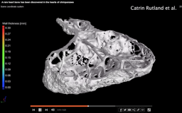
CT images with Dr Catrin Rutland and Dr Craig Sturrock, The Hounsfield Facility, University of Nottingham. |
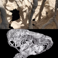
Image Collage. Image by Deborah Gardner. |
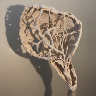
Final artwork by Deborah Gardner, sculpture made from plaster and jesmonite.
Find more about Deborah’s work on her website and on Instagram.
Rachel Rea
Rachel is very satisfied about her collaboration:
'I had great fun working within the group not least because of our distinctly different artistic approaches. It was an honour working with such an eminent scientist as Catrin who communicated her research effectively to support us in our artistic endeavours. The experience has been a stimulating challenge and I would like to continue future collaboration using art to visually communicate scientific subjects.'
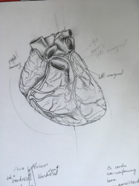
Work in progress. She focused on the understanding of Os Cordis and its location to the heart. Image by Rachel Rea. |
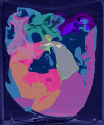
Heart Adaption work in progress. Image by Rachel Rea. |
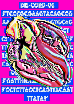
Final artwork by Rachel Rea, illustration, digital print.
She describes her artwork:
'I wanted to take a lateral approach to visually interpret the large amounts of data we received and stay true to the anatomical significance of Catrin's research. I had dissected a Cow's heart in order to better understand what the Os Cordis is and where it is situated within the heart. In this illustration, X marks the spot where the bone can be found, amongst superimposed layers of data that results in a contemporary style.'
Find more about Rachel’s work on her website and on Instagram.
Jess Parry
Jess talks about her inspiration behind her art piece:
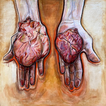
'Catrin described the heart as ‘intimate’ and an ‘emotional heart’ that’s a ‘lifeforce’. From the start, I sensed that Catrin has a deeply personal relationship between her hands and the visceral heart. I always questioned how could I represent this?
'In my collaboration with her, she had initially sent numerous visual materials where I developed a visual understanding of her research. She also delivered a very insightful lecture on her work which helped me understand the context of the visual imagery I was given.
'From this, then as an artist, I had to visually respond to her work. I am a painter so I knew I had to paint something that would strike Catrin on an emotional level and I wanted it to have a heightened sense of realism, to capture the ‘intimate’ and graphic sensuality of such a matter.
'She wanted ‘images to solve the problems that words can’t’ and wanted us to ‘get across the science’.'
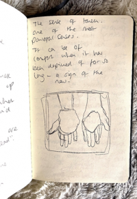
Sketchbook notes. Image by Jess Parry |
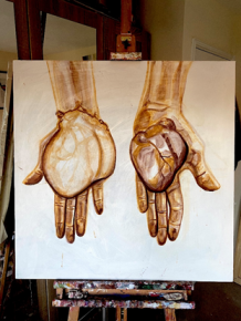
Work in progress. Image by Jess Parry. |
Find more about Jess’s work on her website and on Instagram.
Meetings took place via zoom due to Covid. I had a chance to attend one of those and e-meet the team. The bond and enthusiasm between these 4 women were clear from the start. It is nice seeing three artists of different mediums translating Catrin’s research through the language of art.
Catrin comments about the collaboration:
‘I was so lucky to be able to work with three amazing artists. Seeing what inspired them, their very different takes on my research, and the brilliant ways they were able to interpret science really opened up my eyes. It also interested me to see the way they used my language and how I spoke about the science. It seemed that each of them were driven by particular phrases I used, it reminded me of the power of language between art and science.’
Jess continues:
‘It’s a shame that we as a team haven’t met yet in the flesh, but that is something to look forward to in the future over quite literally ‘a pint of science’.’
No one knows how each of the artist’s career will unfold in the future or if they manage to meet but I hope they will look back to this experience with a smile, as they have achieved such a great body of work regardless of these crazy times of the pandemic.
About this post
This blog post is a part of the Creative Reactions Pint of Science 2021 online exhibition. Throughout May and June, we will open the gallery for different examples of collaborations between art and science at creativereactions.com. Find @creativerxns on Twitter, Instagram and Facebook. Find more Creative Reactions blog posts.
Ask questions to Creative Reactions artist in our virtual Q&A form.
